Sadegh Lab

In pursuit of CSF homeostasis: tackling hydrocephalus

We study how cerebrospinal fluid moves, what it carries, and how it shapes the developing brain, and we translate that into new treatments for hydrocephalus.

UC Davis Department of Neurological Surgery Shriners Children's Hospital Northern California
Openings for funded students, fellows, and volunteers
Research

Five questions we are working on

We study the dynamics of cerebrospinal fluid (CSF) and its relationships with brain shape and function. Combining these insights with clinical experience, we design cellular and genetic therapies to treat hydrocephalus and other conditions causing intellectual disability.

For families

Hydrocephalus is a build-up of cerebrospinal fluid inside the brain’s ventricles that raises pressure and can injure developing brain tissue. It is treated today with surgery (a shunt or an endoscopic opening, or “ETV”) to redirect the fluid.

The work described here is laboratory research. The gene and cell therapies we study are preclinical and are not available as treatments for patients. For care, please speak with your child’s neurosurgeon or neurologist; for plain-language information, patient support, and current clinical trials, we recommend the Hydrocephalus Association.

Gene therapy for hydrocephalus

Hydrocephalus is not inevitable. We are building gene therapies that strengthen the brain’s own defenses against it.

Read more

People of all ages can develop hydrocephalus and related brain injury. But hydrocephalus is not inevitable. Some patients can escape it, using the brain's natural defense mechanisms of CSF homeostasis. How does that work? How can we tell which person is at risk? How can we augment these defense mechanisms in vulnerable patients?

Gene therapy can target and support the critical brain epithelial cells that work to prevent hydrocephalus. We use advanced imaging tools (MRI), hydrodynamic profiling, and animal models of acquired hydrocephalus to study the optimal conditions for such a therapy to work in patients.

Fluidic neuromodulation

Every neuron is bathed in cerebrospinal fluid. If we change the fluid deliberately, we may be able to change how the brain behaves.

Read more

Every neuron in the brain is bathed in cerebrospinal fluid. Its ions, hormones, growth factors, and enzymes are known to shape how neurons behave, which raises a practical possibility: if the composition of CSF can be changed deliberately, brain activity can be modulated through the fluid rather than through the tissue. We call this fluidic neuromodulation, and neurosurgery is already in a position to attempt it, since we routinely drain, divert, and replace CSF.

To test it we need a readout. Through our neurosurgical biobank we collect CSF from patients, including preterm infants after intraventricular hemorrhage, apply it to cultured human neurons, and measure what happens electrically, such as single-cell currents by patch clamp and network activity across multielectrode arrays, in collaboration with Roy Ben-Shalom and Caren Armstrong in the Department of Neurology. Infant CSF collected soon after a hemorrhage disrupts the synchrony of neuronal firing, and that disruption changes as the hemorrhage resolves.

This gives us a way to approach the question neonatologists and neurosurgeons face most often: whether and when to clear blood from the ventricles of a premature infant. Rather than judging a surgery by ventricular size alone, we ask whether it leaves the developing brain in better electrical health.

Early brain development

The fetal brain builds its cell types and its fluid environments together. We think the tiny choroid plexus has an outsized role.

Read more

There is no better way to identify cures for neurologic disorders than by understanding how the fetal brain develops its diversity of cell types and fluid environments over time. The interaction of CSF with these early cell types is avidly debated, and we hypothesize that the tiny choroid plexus plays an outsized role.

The choroid plexus is a neglected part of the brain. Bearing no resemblance to its neighboring neurons and glia, the choroidal cells hide their common heritage. The same early stem cells that produce the hippocampus also generate the choroidal tissue, although with an entirely different purpose. The choroidal tissue does for the brain what the endocrine system, pancreas, liver, and kidneys do for the rest of the body. And there is a lot more to discover.

Intracranial pressure waveform trace with detected peaks marked

Fluid oscillations in the brain

Brain fluids may not move as one. We are measuring pressure in separate compartments to find out.

Read more

Brain fluids may not move as one. Pressure and flow waveforms recorded in different brain compartments appear to be slightly out of step within a single heartbeat, which would mean the fluid spaces do not share pressure instantaneously.

This hydrodynamic view may explain conditions that pressure alone does not: Chiari malformation and syringomyelia, pseudotumor cerebri, sunken flap syndrome, and the ventriculomegaly seen in astronauts after long spaceflight. It also gives us a way to ask why shunt alternatives such as endoscopic third ventriculostomy work when they do, and to predict which one will work for a given child.

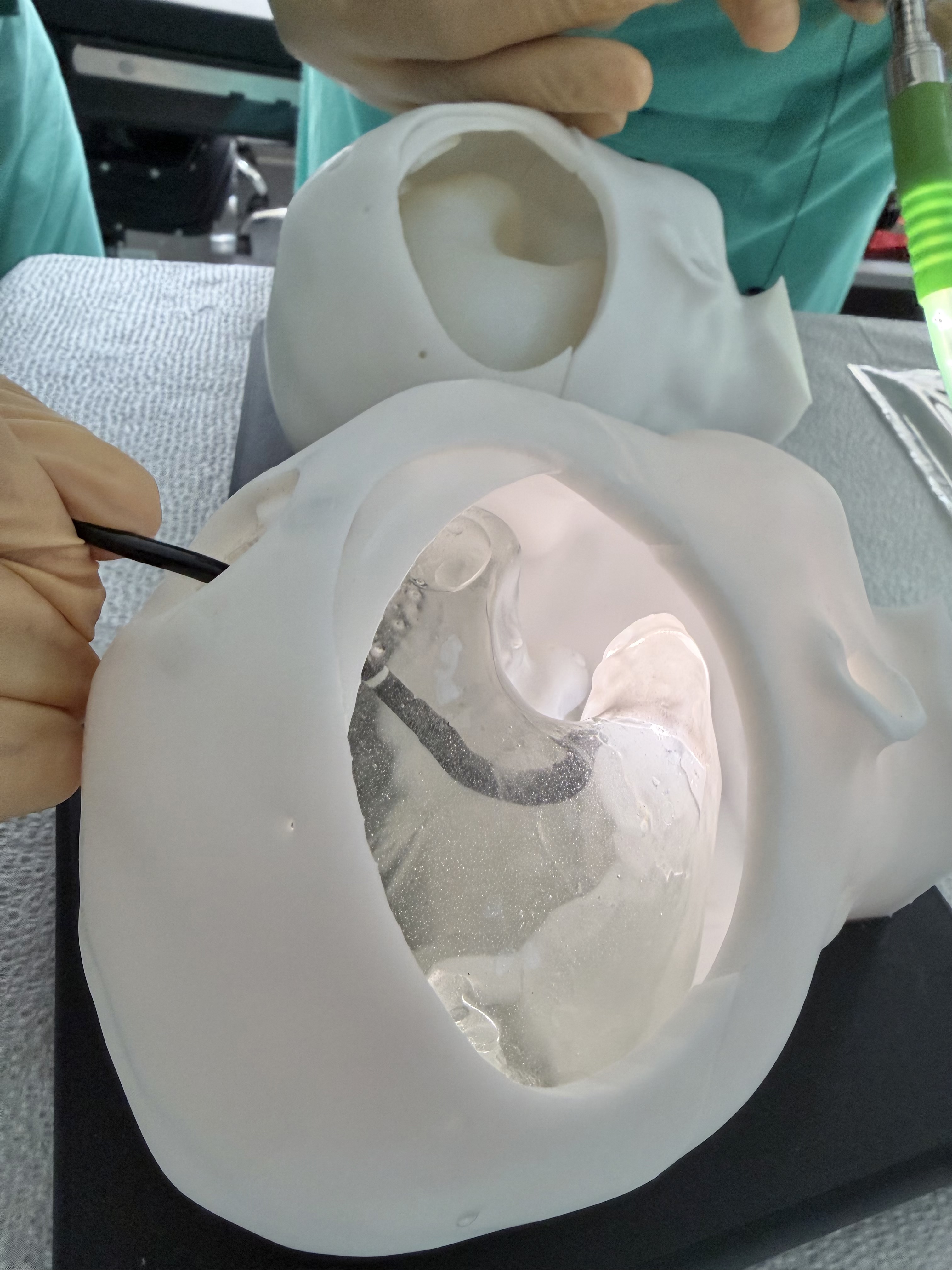
Endoscope entering a transparent 3D printed model of a child’s ventricles

Surgical innovation and One Health

Flexible endoscopy, millimeter-scale robotics, and lessons that travel between children and animals.

Read more

Flexible endoscopy has untapped potential in the fluid spaces of the brain. We develop and test intraoperative technologies there, such as biophotonic imaging, non-contact ablation, and millimeter-scale robotics, with the goal of a genuinely minimally invasive pediatric neurosurgery, alongside 3D printed brain models for training the next generation.

Congenital hydrocephalus occurs naturally in other species, and children and animals can benefit from the same advances. Working with the UC Davis School of Veterinary Medicine, we applied techniques developed for infants to Viggo, a kitten with hydrocephalus, in the first successful endoscopic third ventriculostomy of its kind.

People

The lab

Clinicians, scientists, and students working between the operating room and the bench. Select anyone to read their bio.

Faculty and staff
Undergraduate researchers
Alumni
The Sadegh Lab team

Join the lab

We train students, residents, and visiting scholars at the intersection of CSF physiology, brain development, and pediatric neurosurgery. We are currently able to host trainees who arrive with their own support, and we are glad to help strong candidates compete for it.

Funded students and fellows

Graduate, medical, and postdoctoral trainees holding (or applying for) a fellowship, training grant, or home-institution support. Write early and we will shape the project and the application together.

Undergraduate researchers

UC Davis students can join for research credit or as volunteers, and we support applications to campus and national summer research programs. We ask for a commitment of two years and at least one full summer.

Visiting scholars and clinicians

Short and long-term research visits for clinicians and scientists with external or institutional funding, including international scholars.

On salaried positions: we do not have an open postdoctoral or research specialist line at this time. We do consider exceptional candidates case by case, so if your interests align closely with the lab, please write and include a short note on your interests, a CV, and any funding you hold or plan to apply for.

Publications

Selected work by research area

Choroid plexus and CSF biology

How the choroid plexus builds and clears the fluid the brain develops in.

Neurodevelopment and stem cells

Directing stem cells toward the neuron types lost in injury and disease.

Reviews and field consensus

Setting research priorities for hydrocephalus with the wider community.

Surgical innovation

Augmented reality navigation, flexible endoscopy, and fetal repair.

Clinical reports and outcomes

Cases and outcome studies from pediatric neurosurgical practice.
Collaborators

The work is shared

Our projects run with clinicians and scientists at UC Davis and beyond. Their labs:

More collaborating labs9 more
News

In the press

Support the lab

What your gift does

  • $500 covers the reagents for one month of CSF proteomic analysis from our neurosurgical biobank.
  • $2,500 supports one undergraduate researcher for a summer in the lab.
  • $10,000 funds a pilot experiment testing whether a patient's own CSF changes how human neurons fire, the kind of preliminary result that unlocks federal funding.

Recent support for the lab includes the Hartwell Foundation Individual Biomedical Research Award, the Hydrocephalus Association Innovator Award, the Pediatric Hydrocephalus Foundation, the Chu Family Foundation, the UC Davis Center for Companion Animal Health, EveryCat Health Foundation, and development grants from Shriners Children’s.

Hydrocephalus and Brain Fluid Research Fund

Gifts go directly to lab research and are tax-deductible through UC Davis. Every contribution is genuinely appreciated. Thank you.

Make a gift